Meticulous design very promising
The researchers consider timing a crucial factor for the success of the rehabilitation: An early application of growth stimulators – such as antibodies against the protein Nogo-A – triggers an increased sprouting and growth of nerve fibers. The subsequent training is essential to sift out and stabilize the key neural circuits for the recovery of the motor functions. For instance, an automatic, computer-based analysis of the anatomical data from the imaging revealed that new fibers in the spinal cord sprouted in another pattern depending on the course of treatment. By reversibly deactivating the new nerve fibers that grow, the neurobiologists were ultimately able to demonstrate for the first time that a group of these fibers is essential for the recovery of the motor function observed: Nerve fibers that grew into the spinal cord from the intact front half of the brain – changing sides – can reconnect the spinal cord circuits of the rats' paralyzed limbs to the brain, enabling the animals to grip again.
"Our study reveals how important a meticulous therapeutic design is for the most successful rehabilitation possible," sums up study head Martin Schwab. "The brain has enormous potential for the reorganization and reestablishment of its functions. With the right therapies at the right time, this can be increased in a targeted fashion."
###
A successful interdisciplinary approach
The study was a collaborative project between biologists, physicians and computer scientists. The interdisciplinary team headed by Professor Martin Schwab from UZH's Brain Research Institute and UZH and ETH Zurich's Neuroscience Center included Professor Fritjof Helmchen's group within the same institute and, at the University of Heidelberg, Professor Björn Ommer's group at the Heidelberg Collaboratory for Image Processsing and the Interdisciplinary Center for Scientific Computing.
Literature :
Wahl, A.S., Omlor, W., Rubio, J.C., Chen, J.L., Zheng, H., Schröter, A., Gullo, M., Weinmann, O., Kobayashi, K., Helmchen, F., Ommer, B., Schwab, M.E. Asynchronous therapy restores motor control by rewiring of the rat corticospinal tract after stroke. Science, June 13, 2014. doi: 10.1126/science.1253050
Contact:
Prof. Martin E. Schwab
Brain Research Institute University of Zurich Neuroscience Center Zurich of ETH and University of ZurichWinterthurerstr. 1908057 ZurichPhone: +41 44 635 3330E-mail: schwab@hifo.uzh.ch
Dr. Anna-Sophia Wahl
Brain Research Institute University of Zurich Neuroscience Center Zurich of ETH and University of ZurichWinterthurerstr. 1908057 ZurichPhone: +41 44 635 3228E-mail: wahl@hifo.uzh.ch
Bettina Jakob
Media RelationsUniversity of ZurichTel. +41 44 634 44 39E-mail: bettina.jakob@kommunikation.uzh.ch
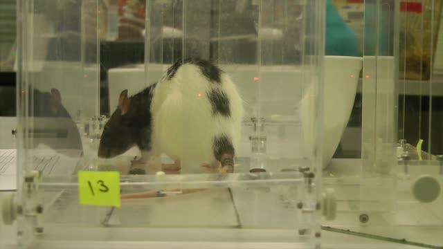
This video depicts Restored grasping after immunotherapy and rehabilitative training.
(Photo Credit: Wahl/Schwab)
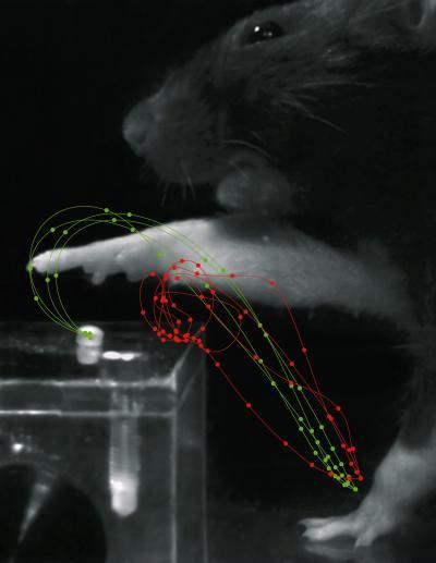
The red trajectories show a rats movements after stroke. Suffering from motor deficits, the rat is unable to control its forelimb function and misses the sugar pellet. After rehabilitation the grasps are smooth and distinct (green trajectories).
(Photo Credit: Tabea Kraus)
Source: University of Zurich