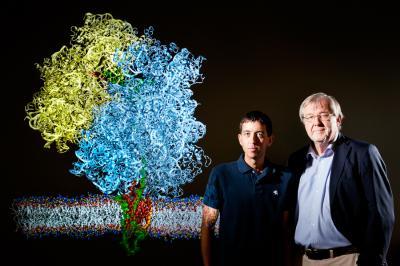
CHAMPAIGN, Ill. — In two new studies, researchers provide the first detailed view of the elaborate chemical and mechanical interactions that allow the ribosome – the cell's protein-building machinery – to insert a growing protein into the cellular membrane.
The first study, in Nature Structural and Molecular Biology, gives an atom-by-atom snapshot of a pivotal stage in the insertion process: the moment just after the ribosome docks to a channel in the membrane and the newly forming protein winds its way into the membrane where it will reside.
A collaboration between computational theoretical scientists at the University of Illinois and experimental scientists at University of Munich made this work possible. Using cryo-electron microscopy to image one moment in the insertion process, the researchers in Munich were able to get a rough picture of how the many individual players – the ribosome, membrane, membrane channel and newly forming protein – come together to get the job done. Each of these structures had been analyzed individually, but no previous studies had succeeded in imaging all of their interactions at once.
"The computational methodology contributed by the Illinois group was crucial in interpreting the new cryo-EM reconstruction in terms of an atomic level structure, and testing the interpretation through simulation," said co-author Roland Beckmann at the University of Munich. "Our joint study is unique in so closely and successfully combining experimental and computational approaches."
To image the ribosome's interaction with the membrane, Beckmann's team used small disks of membrane held together with belts of engineered lipoproteins. University of Illinois biochemistry professor Stephen Sligar developed and pioneered the use of these "nanodiscs."
The Illinois team used the cryo-EM images as well as detailed structural information about the ribosome and other molecules to construct an atom-by-atom model of the whole system and "fit the proteins into the fuzzy images of the electron microscope," said University of Illinois physics and biophysics professor Klaus Schulten, who led this part of the analysis with postdoctoral researcher James Gumbart.

University of Illinois biophysics professor Klaus Schulten (right) and postdoctoral researcher James Gumbart used cryo-EM images as well as detailed structural information about the ribosome and other molecules to construct an atom-by-atom model of the system that threads a growing protein into the cellular membrane.
(Photo Credit: L. Brian Stauffer)
"The ribosome with the membrane and the other components is a simulation of over 3 million atoms," Schulten said, a feat accomplished with powerful computers and "over 20 years of experience developing software for modeling biomolecules." (Schulten is principal investigator of the NIH-funded Resource for Macromolecular Modeling and Bioinformatics at Illinois, which supports the study of large molecular complexes in living cells, with a special focus on the proteins that mediate the exchange of materials and information across biological membranes.)
This analysis found that regions of the membrane channel actually reach into the ribosome exit to help funnel the emerging protein into the channel. Depending on the type of protein being built, the channel will thread it all the way through the membrane to secrete it or, as in this case, open a "side door" that directs the growing protein into the interior of the membrane, Schulten said. The researchers also saw for the first time that the ribosome appears to interact directly with the membrane surface during this process.
The researchers found that a signal sequence at the start of the growing protein threads through the channel and anchors itself in the membrane. Previous studies suggested that this signaling sequence "tells" the ribosome what kind of protein it is building, directing it to its ultimate destination inside or outside the cell.
"This new work visualizes this process for the first time, giving researchers the first image of how nascent proteins actually get into membranes," Schulten said. "It's like going to Mars and being the first to look at Mars."
In a second study, in the Proceedings of the National Academy of Sciences, Schulten, Gumbart and graduate student Christophe Chipot found that proteins get inserted into the membrane in two stages. First, the ribosome "pushes" the growing protein into the membrane channel, and then, in a second step, the protein enters the membrane.
The original push, driven by the chemical energy that the ribosome harvests from other high-energy molecules in the cell, allows even highly charged proteins to pass easily into the oily, nonpolar environment of the membrane, the researchers found.