According to the Agency for Healthcare Research and Quality, more than 300,000 total hip replacements are performed in the United States each year. The procedure reduces pain and restores mobility. However, for younger, more active patients, an artificial hip has a limited lifetime and usually requires restricted activity. Now, researchers at the University of Missouri School of Medicine have tested a new biological technique that may provide better and longer-lasting joint function.
"Resurfacing a joint with donated bone and cartilage tissue is often a better option for young patients with active lifestyles," said Brett Crist, M.D., an associate professor of orthopaedic surgery at the MU School of Medicine and lead author of the study. "Traditional repairs using metal and plastic components begin to wear immediately, so patients must limit activity to reduce damage to their new joints. Although a biological approach may be a better solution, there is no standard method for implantation. Our team compared a common technique with a method developed at the Missouri Orthopaedic Institute to determine if we could further improve joint function."
A common method of implanting donor tissue into the femur part of the hip joint is to use multiple small, cylinder-shaped plugs of bone and cartilage to fill in a damaged area. Crist's team investigated a newer method using larger, size-matched grafts to cover the area in need of repair. The larger grafts also have beveled edges to provide a more precise fit.
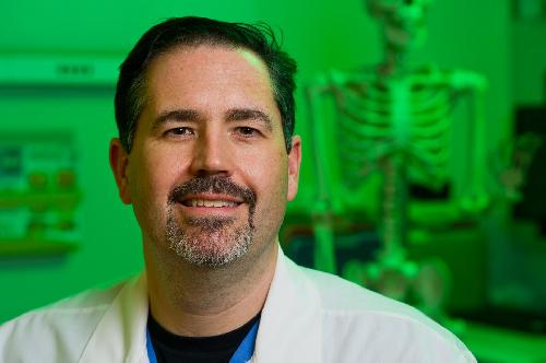
 This is Brett Crist, M.D., associate professor of orthopaedic surgery at the MU School of Medicine and lead author of the study. Credit: Justin Kelley, University of Missouri Health
The researchers used dog femurs to compare small grafts taken from the knee area of the dog to small and large grafts taken from donor dogs. After surgery, the dogs were allowed unrestricted activity and walked on a leash for 15 minutes, five times a week.
The researchers found that the dogs implanted with traditional small grafts showed significant loss in range of motion and joint integrity after only eight weeks. In contrast, the dogs implanted with larger, bevel-shaped grafts maintained joint viability and structural integrity throughout the six-month study period.
"By using one large graft, we reduced the number of seams for a smoother functioning joint," Crist said. "Beveling the edges also created a better fitting repair that was less prone to cell death during implantation."
More studies are needed to verify the optimal size and technique for implanting donor grafts in the hip. However, the study provides initial clinical evidence that larger, size-matched grafts have the potential to improve outcomes when resurfacing cartilage defects of the femoral head in the hip joint.
The study, "Optimizing Femoral-head Osteochondral Allograft Transplantation in a Preclinical Model," recently was published in the Journal of Orthopaedic Translation.
source: University of Missouri-Columbia