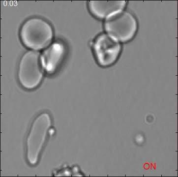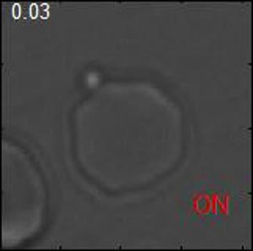
This video shows the delivery of a viable merozoite via optical tweezers to a healthy erythrocyte and subsequent invasion.
(Photo Credit: Biophysical Journal, Crick et al.)

This video shows local erythrocyte membrane deformations induced by tweezer-manipulation of post-viable merozoite contact area.
(Photo Credit: Biophysical Journal, Crick et al.)

This video shows the forcible detachment of an adherent merozoite from the erythrocyte surface via optical tweezers.
(Photo Credit: Biophysical Journal, Crick et al.)
Source: Cell Press