Smartphones are capable of giving us directions when we're lost, sending photos and videos to our friends in mere seconds, and even helping us find the best burger joint in a three-mile radius. But University of Houston researchers are using smartphones for another very important function: diagnosing diseases in real time.
The researchers are developing a disease diagnostic system that offers results that could be read using only a smartphone and a $20 lens attachment.
The system is the brainchild of Jiming Bao, assistant professor of electrical and computer engineering, and Richard Willson, Huffington-Woestemeyer Professor of Chemical and Biomolecular Engineering. It was created through grants from the National Institutes of Health and The Welch Foundation, and was featured in February in ACS Photonics.
This new device, like essentially all diagnostic tools, relies on specific chemical interactions that form between something that causes a disease – a virus or bacteria, for example – and a molecule that bonds with that one thing only, like a disease-fighting antibody. A bond that forms between a strep bacteria and an antibody that interacts only with strep, for instance, can support an ironclad diagnosis.
The trick is finding a way to detect these chemical interactions quickly, cheaply and easily. The solution proposed by Bao and Willson involves a simple glass slide and a thin film of gold with thousands of holes poked in it.
Creating this slide is itself an achievement. This task, led by Bao, starts with a standard slide covered in a light-sensitive material known as a photoresist. He next uses a laser to create a series of interference fringes – basically lines – on the slide, and then rotates it 90 degrees and creates another series of interference fringes. The intersections of these two sets of lines creates a fishnet pattern of UV exposure on the photoresist. The photoresist is then developed and washed away.
While most of the slide is then cleared, the spots surrounded by intersecting laser lines – the 'holes' in the fishnet – remain covered, basically forming pillars of photoresist.
Next, he exposes the slide to evaporated gold, which attaches to photoresist and the surrounding clean glass surface. Bao then performs a procedure called lift-off, which essentially washes away the photoresist pillars and the gold film attached to them.
The end result is a glass slide covered by a film of gold with ordered rows and columns of transparent holes where light can pass through.

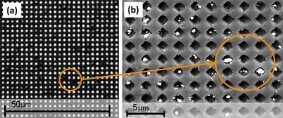
LEFT: The system being developed by UH Cullen College of Engineering researchers diagnoses disease by blocking holes with pathogens and some other connected material, in this case silver particles, preventing light from shining through. RIGHT: This is a close-up of nanoholes blocked by these particles.
(Photo Credit: Jiming Bao Research Group)
These holes, measuring about 600 nanometers each, are key to the system. Willson and Bao's device diagnoses an illness by blocking the light with a disease-antibody bond – plus a few additional ingredients.
Here is where Willson comes in. An internationally known biomolecular engineer, Willson starts by placing disease antibodies in the holes, where they are coaxed into sticking to the glass surface. Next, he flows a biological sample over the slide. If the sample contains the bacteria or virus being sought out, it will bond with the antibody in the hole.
This bond alone, though, doesn't block the light. "The thing that binds to the antibody is probably not big and grey enough to darken this hole, so you have to find a way to darken it up somehow," Willson said.
Willson achieves this by flowing a second round of antibodies that bond with the bacteria over the slide. Attached to these antibodies are enzymes that produce silver particles when exposed to certain chemicals. With this second set of antibodies now attached to any bacteria in the holes, Willson then exposes the entire system to the chemicals that encourage silver production.
About 15 minutes later he rinses off the slide. Thanks to chemical properties of the gold, the silver particles in the holes will remain in place, completely blocking light.
Here's where the smart phone comes in. One of the advantages of this system is that the results can be read with simple tools. A basic microscope used in elementary school classrooms, Willson said, provides enough light and magnification to show whether the holes are blocked. With a few small tweaks, a similar reading could almost certainly be made with a phone's camera, flash and an attachable lens.
This system, then, promises readouts that are affordable and easy to interpret.
"Some of the more advanced diagnostic systems need $200,000 worth of instrumentation to read the results," said Willson. "With this, you can add $20 to a phone you already have and you're done."
There are still major technical hurdles to clear before the system can be rolled out, Willson noted. One of the biggest challenges is finding a way to drive the bacteria and viruses in the sample down to the surface of the slide to ensure the most accurate results.
But if those problems are overcome, the system would be an excellent tool for healthcare providers in the field.
At the site of an industrial accident, for instance, the holes on a single slide could be populated with molecules that bond with 10 potential contaminants, allowing response teams to quickly assess the situation. In economically disadvantaged areas, such a system could be used to screen large groups of people for widespread and serious health problems, like diabetes.
"There are a lot of situations where an affordable diagnostic tool that is simple to use and simple to interpret could be very useful," said Willson. "If both your disposables and your reader are cheap, that makes it a lot easier to extend your system out into the real world."
Source: University of Houston