OAK BROOK, Ill. – Incidental chest computed tomography (CT) findings can help identify individuals at risk for future heart attacks and other cardiovascular events, according to a new study published online in the journal Radiology.
"In addition to diagnostic purposes, chest CT can be used for the prediction of cardiovascular disease," said Pushpa M. Jairam, M.D., Ph.D., from the University Medical Center Utrecht, in Utrecht, the Netherlands. "With this study, we have taken a new perspective by providing a different approach for cardiovascular disease risk prediction strictly based on information readily available to the radiologist."
Currently, individuals at high risk for cardiovascular events are identified through risk stratification tools based on conventional risk factors, such as age, gender, blood pressure, cholestorol levels, diabetes, smoking status or other factors thought to be related to heart disease. However, a substantial number of cardiovascular events occur in individuals with no conventional risk factors, or in patients with undetected or underdiagnosed risk factors.
"Extensive literature has clearly documented the uncertainty of prediction models based on conventional risk factors," Dr. Jairam said. "With this study, we address to some extent, the need for a shift in cardiovascular risk assessment from conventional risk factors to direct measures of subclinical atherosclerosis."
Through the use of chest CT, radiologists are routinely confronted with findings that are unsuspected or unrelated to the CT indication, known as incidental findings. Incidental findings indicating early signs of atherosclerosis are quite common and could play a role in a population-based screening approach to identify individuals at high risk for cardiovascular events. However, there is currently no guidance on how to weigh these findings in routine practice.

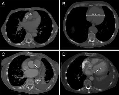
Here are examples of cardiovascular chest CT findings. A, Ascending thoracic aorta diameter measurement.B, Cardiac diameter measurement. C, Calcifications in the left anterior descending coronary artery andthe descending thoracic aorta (arrows). D, Calcifications on the mitral valve (arrow).
(Photo Credit: Radiological Society of North America)
Dr. Jairam and colleagues set out to develop and validate an imaging-based prediction model to more accurately assess the contribution of incidental findings on chest CT in detecting patients at high risk for cardiovascular disease.
The retrospective study looked at follow-up data from 10,410 patients who underwent diagnostic chest CT for non-cardiovascular indications. During a mean follow-up period of 3.7 years, 1,148 cardiovascular events occurred among these patients.
CT scans from these patients and from a random sampling of 10 percent of the remaining patients in the group were visually graded for several cardiovascular findings. The final prediction model included age, gender, CT indication, left anterior descending coronary artery calcifications, mitral valve calcifications, descending aorta calcifications and cardiac diameter. The model was found to have accurately placed individuals into clinically relevant risk categories.
The results showed that radiologic information may complement standard clinical strategies in cardiovascular risk screening and may improve diagnosis and treatment in eligible patients.
"Our study provides a novel strategy to detect patients at high risk for cardiovascular disease, irrespective of the conventional risk factor status, based on freely available incidental information from a routine diagnostic chest CT," Dr. Jairam said. "The resulting prediction rule may be used to assist clinicians to refer these patients for timely preventive cardiovascular risk management."
Dr. Jairam cautions that a prospective, multicenter trial is needed to validate the impact of these findings.