HOUSTON -- (March 21, 2011) -- In a surprising new study, researchers using image-analysis methods similar to those employed in facial-recognition software have made a startling discovery that rules out the two main theories scientists had created to explain how bacteria self-organize into multicellular aggregate mounds. The study by researchers from Rice University and the University of Georgia appears online this week in the Proceedings of the National Academy of Sciences.
The find is important for the study of biofilms -- slimy colonies of bacteria that form on everything from teeth to pacemakers. Federal health officials have estimated that as many as 80 percent of all microbial infections arise from biofilms, and scientists know that the same bacteria can be up to 1,000 times more resistant to antibiotics if they're living inside a biofilm rather than living on their own. To better fight biofilms, scientists have been scrambling to understand the biochemical and biophysical mechanisms that allow bacteria to form aggregates, reorganize and interact.
"The results of our analysis were really surprising," said study co-author Oleg Igoshin, assistant professor in bioengineering at Rice. "Our results didn't support either of the major competing theories people have come up with. Those theories were each predicated on the idea that as the bacterial mounds were forming and reorganizing, the individual bacterium were drawn toward one or another of them by some sort of chemical signal.
"That doesn't appear to be the case at all," Igoshin said. "We didn't find any neighbor-related factors between the groups at all. Instead, there seems to be a signaling mechanism within the group itself that trumps everything else."
The study involved the bacterium Myxococcus xanthus, a common soil bacteria that's often studied for its ability to self-organize into various patterns. In the wild, M. xanthus are content to collectively hunt other bacteria. But when food is scarce, they stream together into aggregates containing up to 100,000 cells and form spores. The resulting aggregate mounds are large enough to be carried away to better environs by the wind or passing insects.

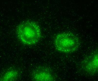
When food is scarce, Myxococcus xanthus bacteria stream together by the thousands to form aggregate mounds.
(Photo Credit: Swapna Bhat and Ricky Patel/Shimkets Lab/University of Georgia)
To study this behavior in the lab, Igoshin and Rice co-authors postdoctoral fellow Chunyan Xie and graduate student Haiyang Zhang created a computer program that could analyze thousands of still frames from microcinematic movies of M. xanthus. The movies were created in the laboratory of University of Georgia collaborator and co-author Lawrence Shimkets. The movies showed how M. xanthus streamed together to form "aggregates." One hallmark of the M. xanthus streaming process is that less than half of the aggregates that initially form will survive through the end of the process. The factors that control this ripening are not understood.
In designing their image-analysis application, Igoshin's team had the computer scrutinize every aggregate -- frame-by-frame -- throughout the streaming process. The computer cataloged 33 properties for each aggregate, including things like area, perimeter size,.distance to and size of the nearest neighbor. After all the data were collected, the team ran a statistical analysis to find out if any feature or combination of features could be used to predict which aggregates would eventually win out over their neighbors.
"We found that size mattered most," Igoshin said. "Not size in relation to neighbors, which is something people had previously thought might matter, but size of the aggregate itself. We found that if we answered one question -- is the size of an aggregate beyond a certain threshold -- then we could accurately predict whether the aggregate would survive with 90 percent accuracy."
Igoshin said some of the image analysis methodologies that the team applied to study M. xanthus are similar to ones that Chunyan Xies used for facial recognition analyses in her previous work. He said scientists have only recently begun to apply these sorts of image analysis techniques to fundamental biological questions like bacterial self-organization.
"One of the most exciting aspects of this study is the fact that we can apply these methods much more broadly to study self-organization in other bacteria and unicellular organisms," Igoshin said. "In fact, this kind of analysis is sorely needed, because most of the existing methods to study these phenomena are qualitative rather than quantitative. As a discipline, we need quantitative methods if we want to conduct side-by-side comparisons between real-world and computer-generated results."
Source: Rice University