For years, the best that lung anatomy pioneers such as study co-corresponding author Ewald Weibel, professor emeritus of anatomy at the University of Bern, could do to study specific areas of a lung was to make measurements in two dimensions or create 3D casts of a lung's air spaces. The techniques, while giving the earliest insights into a lungs's makeup and functioning, had their limitations. For one, they did not directly replicate a lung's structure in real life, and they could not convey how various parts act together as a whole. Yet advances in imaging and computation have enabled researchers to more fully explore how gases and other inhaled substances act in the lung's furthest recesses.
In this study, the team worked with 22 pulmonary acini culled from young and old mice. They then set to "reconstruct" the acini based on micro computed tomography imaging of scanned lungs in mice and extracted from them. The extracted lungs were preserved in a way that kept the anatomy intact—including the tiny air spaces required for successful imaging. From that, the researchers were able to measure an acinus, estimate the number of acini for each mouse lung and even count the alveoli and measure their surface area.
The mouse lung, in its structure and function, is remarkably similar to the human lung. That means researchers can alter the genetics of a mouse and see how those changes affect the peripheral structure of the lung and its performance.
Already, the researchers found in the current study that mouse alveoli increase in number long past the two weeks that at least one previous study had indicated. Hoffman adds that a separate study is needed to determine whether humans, too, increase the number of air sacs past a certain, predetermined age.
The researchers next aim to use the model to more fully understand how gases interact with the bloodstream within the acini and the alveoli.
"Our imaging and image-analysis methodologies enable new ways to investigate the lung's structure and can now be used to further investigate the normal healthy-lung anatomy in humans and be used to visualize and assess the pathological changes in animal models of specific structural diseases," says Vasilescu, who is a postdoctoral researchfellow at the University of British Columbia.
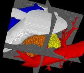
The video shows the imaging of a section of a mouse lung. As the image rotates, more respiratory branches (bronchioles) are shown, along with three acini (yellow, green and orange clusters). The blood vessels feeding the acini are then added with the arteries shown in blue and the veins in red.
(Photo Credit: University of Iowa)
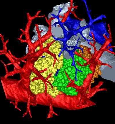
A research team led by the University of Iowa has created the most detailed, three-dimensional rendering of a mammal lung. The image pictured here shows a mouse's pulmonary acini, the terminals where gases and blood mix in a lung and whose function remains a mystery.
(Photo Credit: Dragos Vasilescu, University of Iowa and the University of British Columbia.)
Source: University of Iowa